Back to store
Price: $47,300
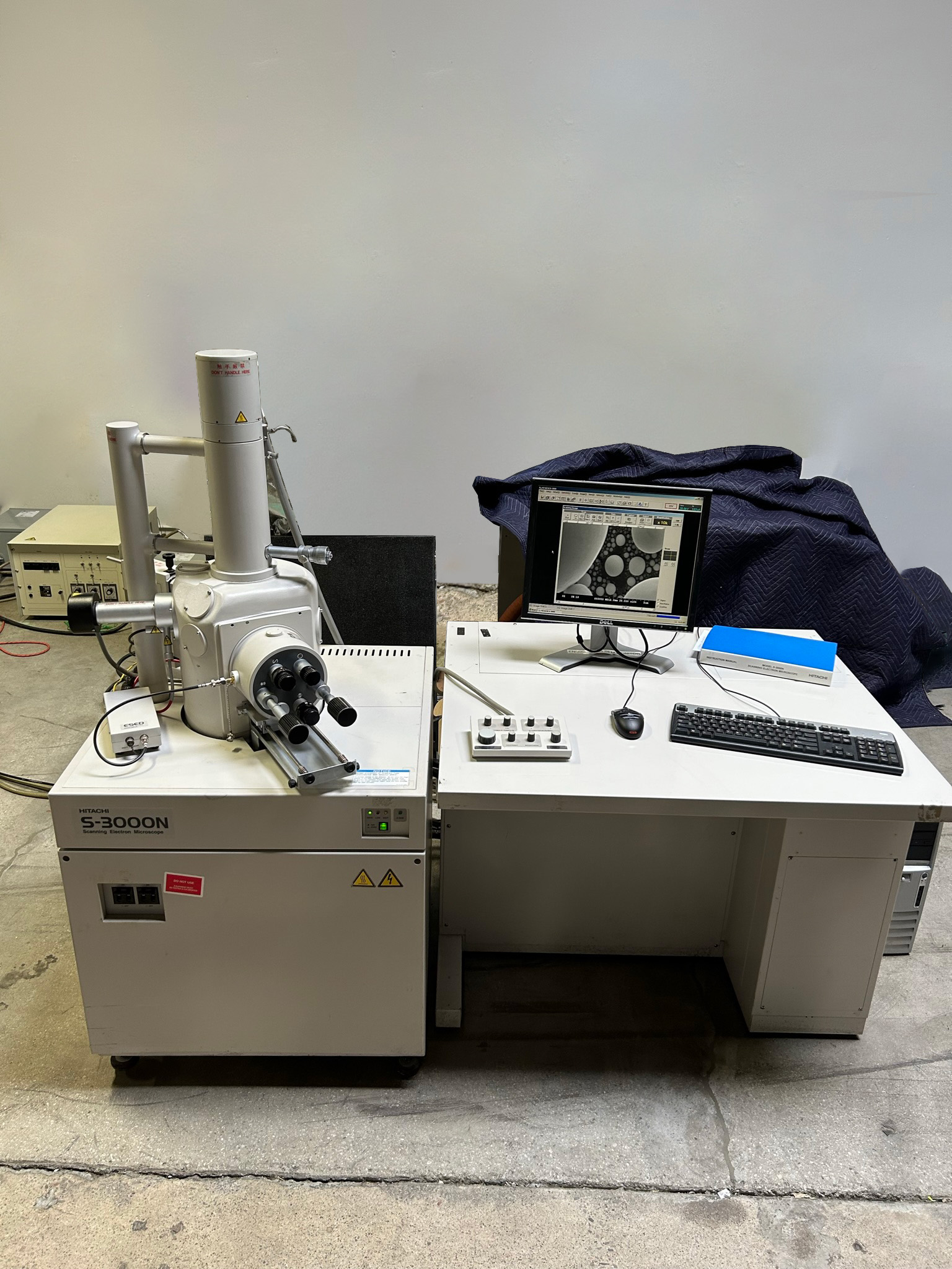
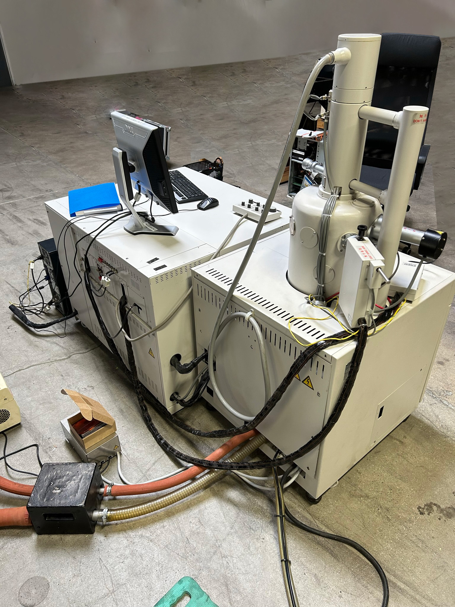
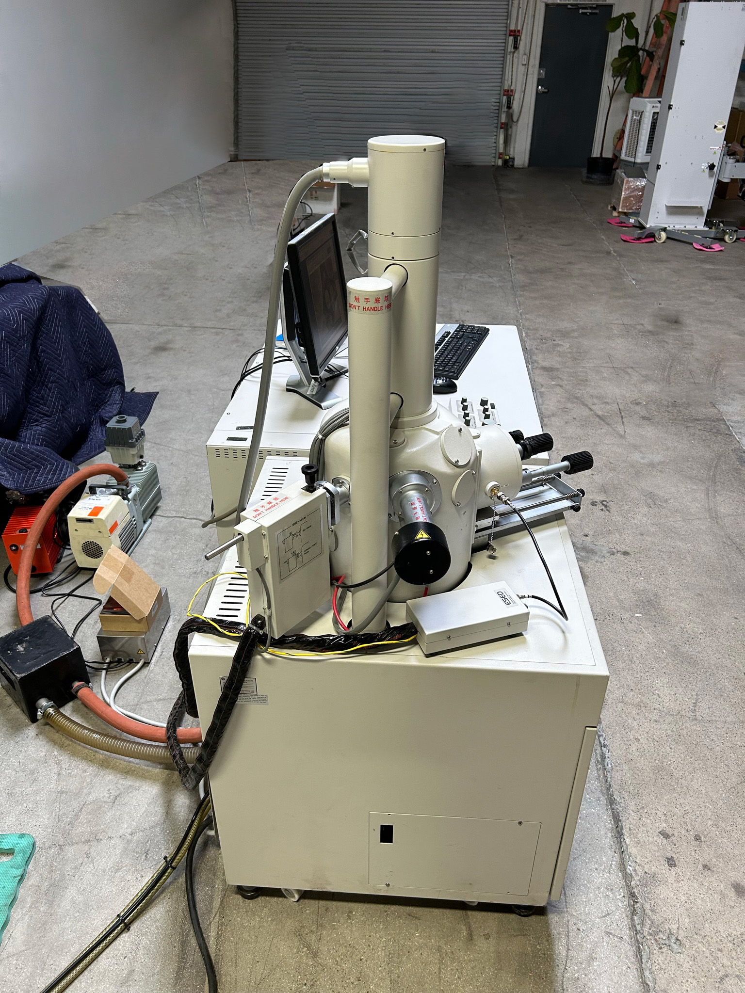
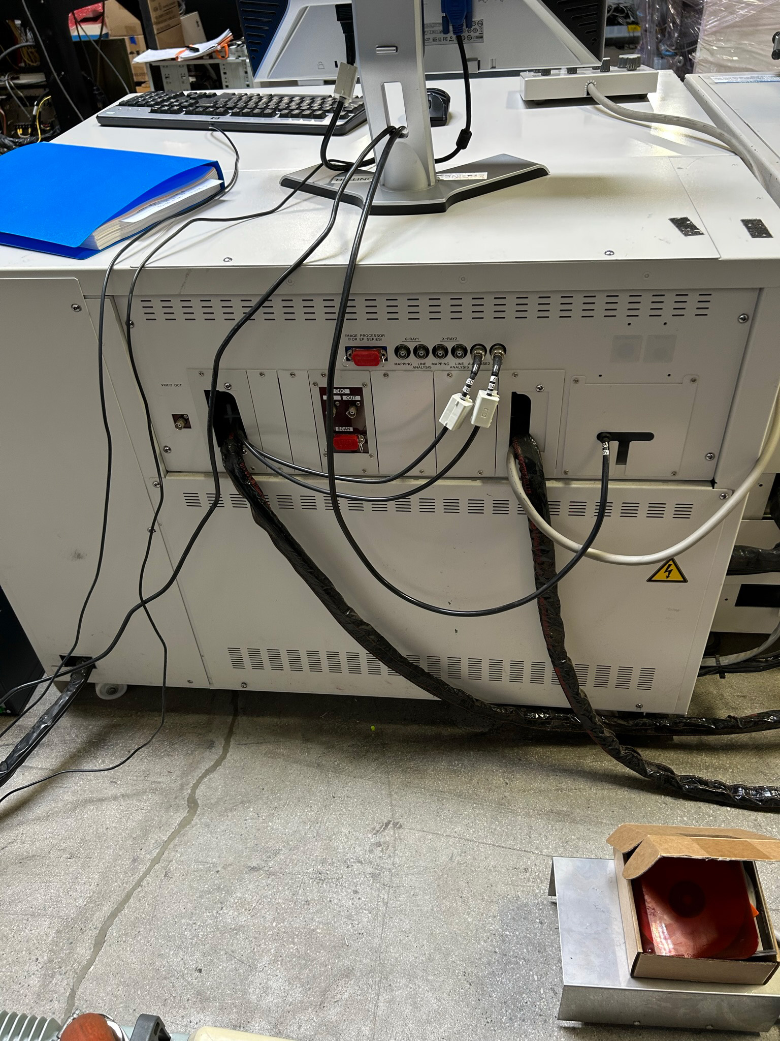
Hitachi S 3000 N Scanning Electron Microscope (SEM) -68534
Price: $47,300
- Asset # : 68534
- Make : Hitachi
- Model : S 3000 N
- Type : Scanning Electron Microscope (SEM)
- Wafer Size :
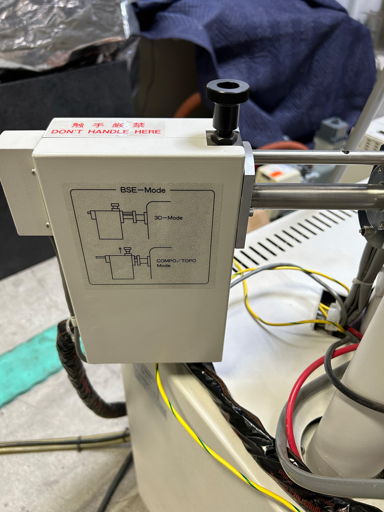
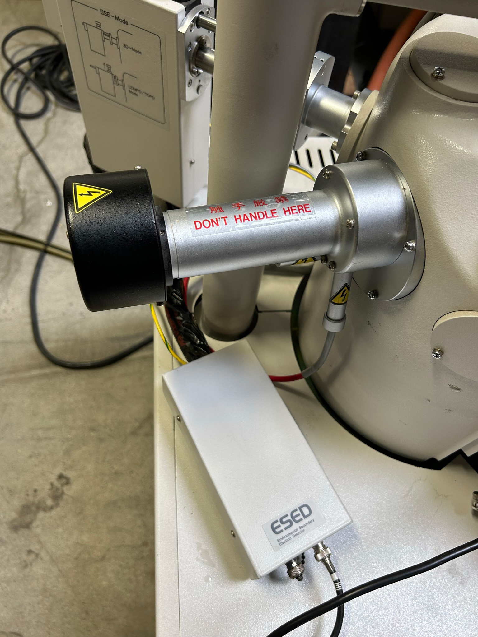
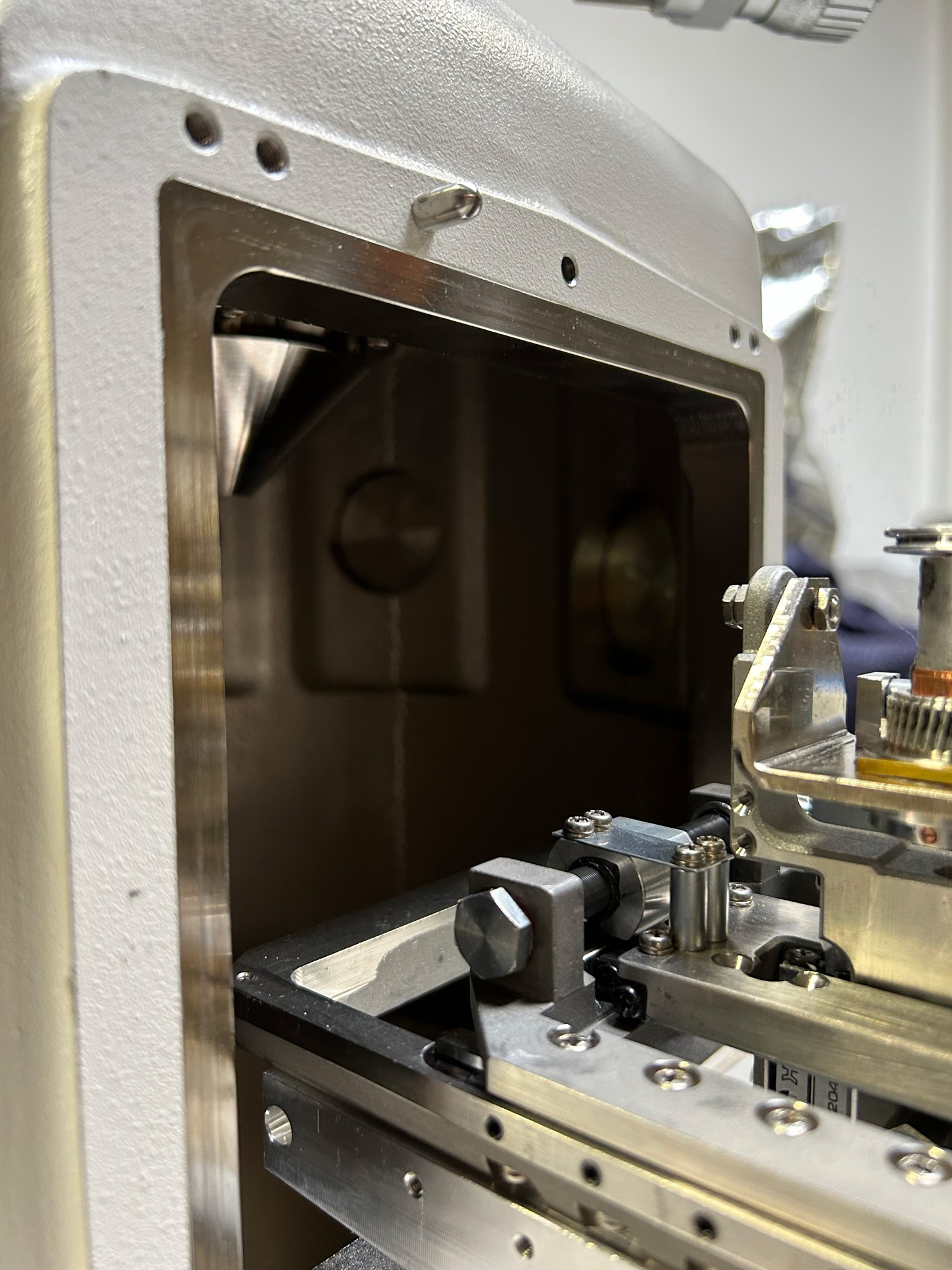
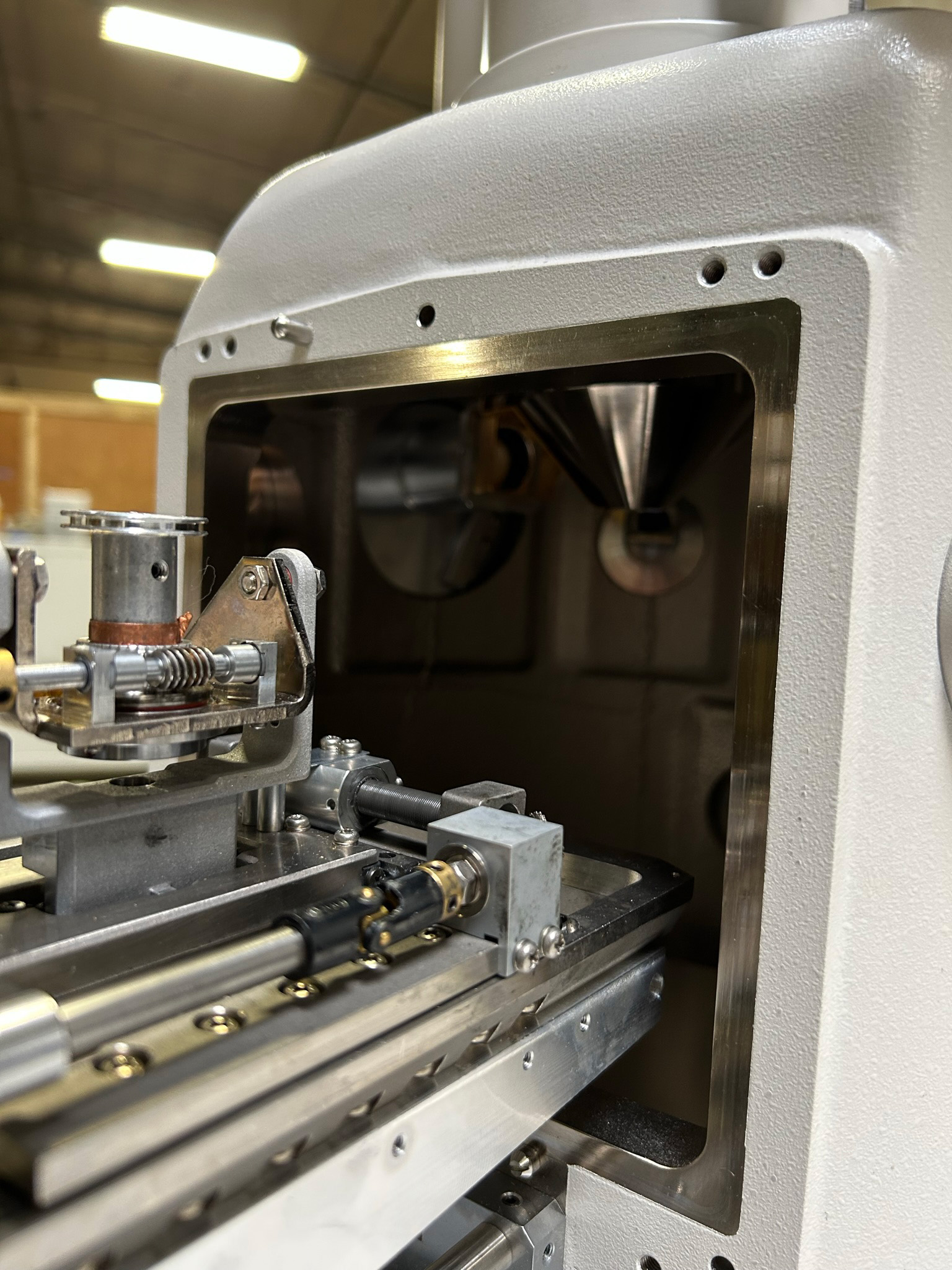
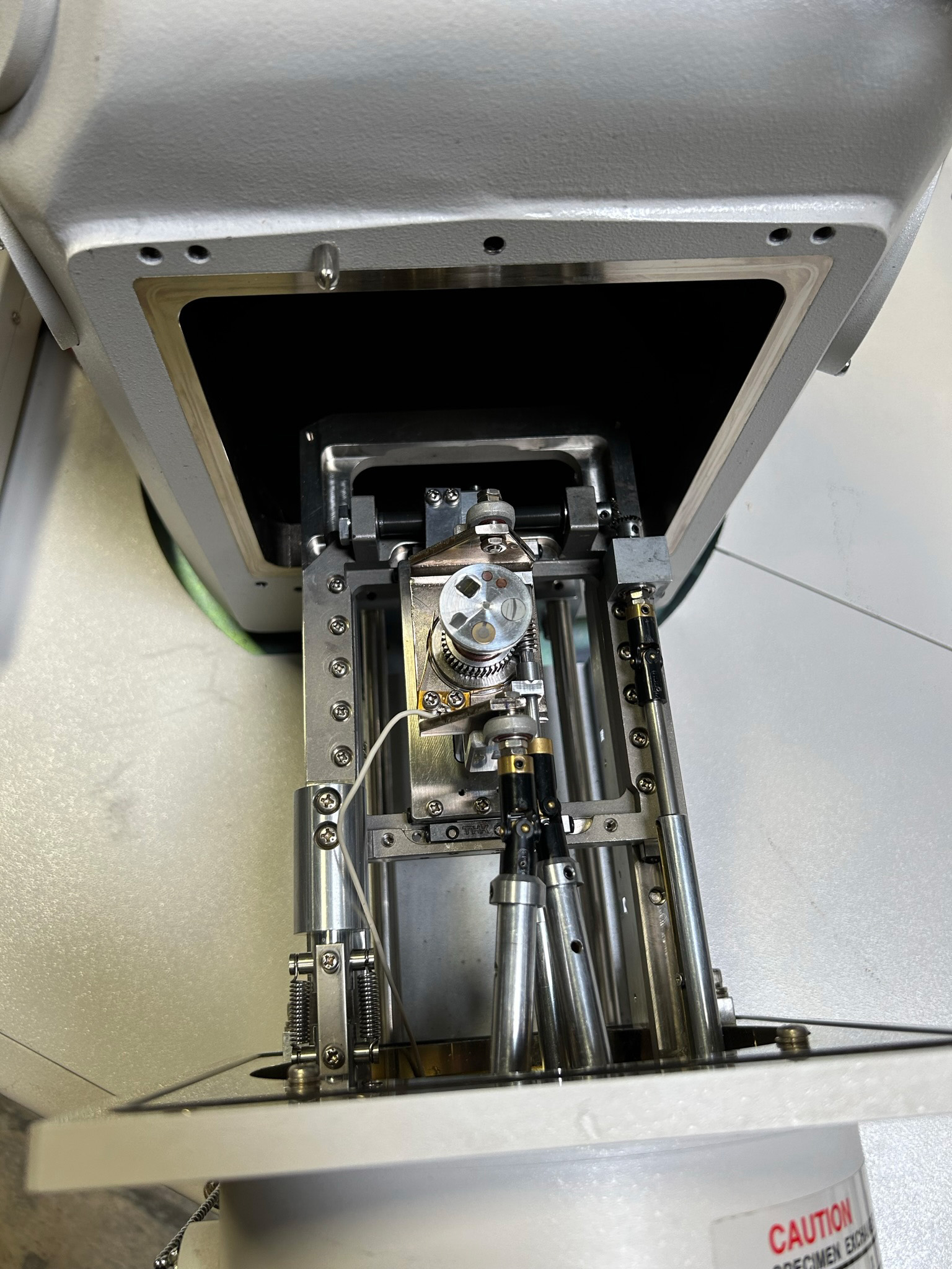
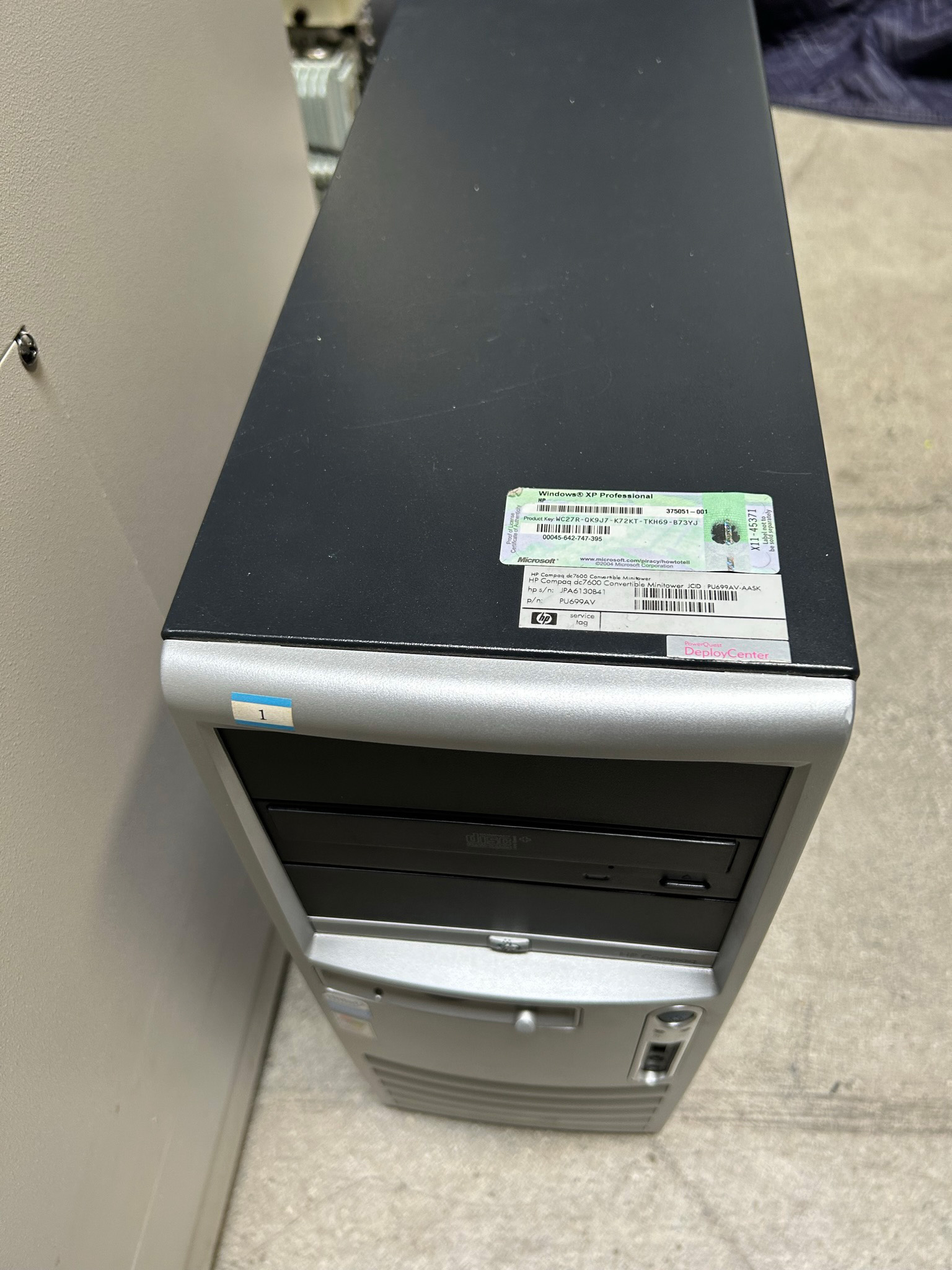
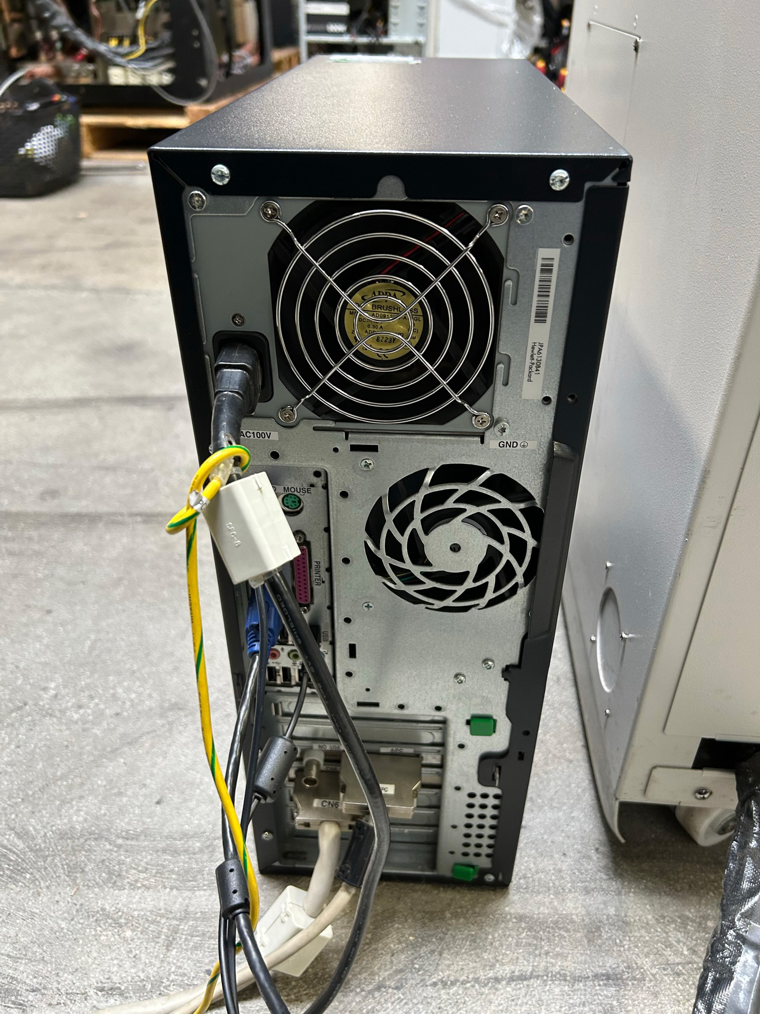
- Configuration : - Variable Pressure (Low Vacuum) - 6" Stage - Windows XP Operating System - HP DC 7600 - Software Options: - Measurement - PCI Interface (Image management) - H.R. Image Memory - SEM Data Management - Full Screen - 5 Axis Stage - 4 Quadrant Backscatter Detector - ESED (AED) Advanced Detector - Option to add EDX for an additional cost Specifications: 1. Resolution: - Secondary electron image resolution: 3.0 nm (High Vacuum Mode) 15 nm (Accelerating voltage 3kV) 2. Magnification: - 15x to 300,000x (65 steps) Normal mode - 5x to 300,000x (75 steps) Low mag mode -(x5 : WD = 35 mm, Accelerating voltage 2kV) 3, Electron Optics: (1) Filament: Pre-centered tungsten hairpin type (2) Gun bias: Self-bias + continuously variable bias (3) Accelerating voltage: -0.3 to 30 kV (1171 steps) -0.3 to 9.99 kV (in increments of 10 V) -10 to 30 kV (in increments of 0.1 kV) (4) Emission current: 10^-12 to 10^-7 A (5) Gun Alignment: 2-stage electromagnetic alignment (6) Condenser lens: 2-stage electromagnetic condenser (7) Objective lens: Super-conical lense (8) Objective lens aperture: 4-opening movable aperture (9) Stigmator coil: 8-pole electromagnetic X/Y correction for astigmatism (10) Image shift: ± 20 μm or more (W.D. = 15 mm) 4. Specimen Stage One of the following five types of specimen stages is selectable. (a) Super-eucentric stage Movement range: 32 mm x 32 mm Tilt angle: -90° to +90° Rotation angle: 360° (b) Standard stage Movement range: 80 mm x 40 mm Tilt angle: -20° to +90° Rotation angle: 360° (c) Large-size eucentric stage Movement range: 100 mm x 50 mm Tilt angle: 0° to +60° Rotation angle: 360° (d) Cool stage 20 Movement range: 15 mm x 15 mm Tilt angle: -45° to +45° Temperature range: +10° C to -20° C (e) 5-axis stage Movement range: 100 mm x 50 mm Tilt angle: 0° to +60° Rotation angle: 360° 5. Image Display (1) Kinds of images: - Secondary electron image (only in HIGH VACUUM mode) - Back-scattered electron image (Semiconductor method) (2) Scanning modes - TV scan - Slow scan (4 steps) - Selected area scan - Waveform monitor/signal monitor - Photo scan (4 steps) - Twin photo scan - Dual-magnification scan (3) Scanning speeds - TV scan - Slow 1: 0.35 s (X = 0.7 ms, Y = 480 lines) - Slow 2: 2 s (X = 4 ms, Y = 480 lines) - Slow 3: 10/8 s (X = 20/16.7 ms, Y = 480 lines)* - Slow 4: 20/24 s (X = 40/50 ms, Y = 480 lines)* - Selected area: 70 ms (F) (X = 0.45 ms, Y = 160 lines One-stage filter) 70 ms (M) (X = 0.45 ms, Y = 16- lines, Two stage filter) 320 ms (S) (X = 2 ms, Y = 160 lines) - Photo 1: 40/33 s (X = 20/16.7 ms, Y = 1920 lines)* - Photo 2: 80/100 s (X = 40/50 ms, Y = 1920 lines)* - Photo 3: 200/200 s (X = 100/100 ms, Y = 1920 lines)* - Photo 4: 400/400 s (X = 200/200 ms, Y = 1920 lines)* (*: Synchonized with power frequency of 50/60 Hz) (4) Signal processing (analog operation Image polarity reversal Image differentiation Gamma Control (5) Data display: Accelerating voltage, magnification, micron scale, micron value, film number, W.D. value, date/time, vacuum level, photo magnification, detector (6) Data entry: Input through full keyboard (alphabetic characters, numerics, symbols), Graphic input (straight lines, circles, arrows, etc.) (7) VTR signal input 6. Image memory (1) For display (640 x 480) (2) For high resolution (1280 x 960) (3) For ultra-high resolution (2560 x 1920) (Option) (4) Memory functions: Scan conversion, recursive filter (applicable modes: TV, Slow 1, slow 2), image integration (2 to 1024 images) (Applicable modes: TV, Slow 1 to Slow 4), Brightness conversion (LUT: Look-up table method), Intermediate level emphasis, gamma control, N-ary conversion, Pseudo color, Histogram presentation, 4-split screen presentation 7. Automatic Functions - Auto brightness and contrast (ABC) - Auto stigmatism and focus (ASF) - Auto focus (AFC) - Auto filament saturation (AFS) - Auto gun alignment (AGA) - Auto start (HV-ON-> AFC-> ABC) - Auto photographing (1. AFC -> ABC-> Photo) (2. ABC -> Photo) - Full auto mode (1: AFS -> AGA-> AFC -> ABC) (2: AGA-> AFC -> ABC) 8. Operation Support Functions - Operation principle: Graphical user interface / simplified menus - Control devices: Mouse, dedicated rotary knob, full keyboard - Magnification presetting (Arbitrary magnification settable) - Axial alignment wobbler - Condition registration (15 conditions, to be registered unlimitedly) - Focus search - Focus memory - ABC Brightness setting - Film sensitivity compensation - Photo magnification compensation - Photo-only brightness setting - Scan point indication - Filament image - Status list indication 9. X-Ray Analysis Mode - Signal input terminals (equipped for each of two systems), X-ray rate meter signal terminal (0 to +10 V), Mapping signal terminal (TTL) - Scan mode: Line analysis, spot analysis, selected area analysis - Analysis position: W.D = 15 mm, X-ray take-off angle (TOA) = 35° 10. Vacuum System: - Principle: Full-auto pneumatic valve control evacuation - Vacuum gauge: Pirani Gauge - Indications: LED, menus - Ultimate vacuum: 1.5 x 10^-3 Pa - Variable pressure mode: Variable pressure range: 1 to 270 Pa, Vacuum level setting: Selectable from 25 steps via dialog - Vacuum pumps: Oil diffusion pump (DP): 570 L/s for high vacuum, oil rotary pump 1 (RP 1): 135 (162) L/min, for DP back pressure evacuation only, Oil rotary pump 2 (RP 2): 135 (162) L/min, for pre-evacuation/low-vacuum setting 11. Protective devices: Protection against power outage and water supply interruption 12. Standard System Composition: - Column unit (1) - Display unit (1) - PC unit (1) - Oil rotary pump (2) - Compressor (1) - Standard tools (1 set) - Spare and consumable parts (1 set) - Instruction manual (1 copy)
Hitachi S 3000 N Scanning Electron Microscope (SEM) -68534
$47,300.00
